XanTec bioanalytics GmbH

Interested in our SPR instruments? Contact us for price information or a quote!

Products | SPR Biosensors | SPR Systems | SPR-PLUS | Applications


*** The NEW SPR-PLUS concept *** The NEW SPR-PLUS concept *** The NEW SPR-PLUS concept ***

Application
option 01*

A novel Method to detect IgG variants: Combined preparative SPR-PLUS and mass spectrometry in FcRn affinity chromatography

Preserving the chemical and structural integrity of therapeutic antibodies during manufacturing and storage is a major challenge during pharmaceutical development. Oxidation of Fc methionines Met252 and Met428 is frequently observed, which leads to reduced affinity to FcRn and faster plasma clearance if present at high levels. Because oxidation occurs in both positions simultaneously, their individual contribution to the concomitant changes in pharmacokinetic properties has not been clearly established1.

We present a novel approach for research into this problem. A new preparative SPR-PLUS system has been used to study FcRn interaction analysis. This system allowed us to isolate antibody oxidation variants from an oxidized IgG1 preparation based on their FcRn binding properties monitored by SPR, fractionized in its dissociation phase and subsequently analyzed by mass spectrometry.

Figure 1
Figure 1: Setup of the SPR-PLUS system for FcRn-chromatography. From left to right: Autosampling unit, pneumatic gradient pump, 2SPR spectrometer, valve manifold, fraction collector.

Materials & Methods

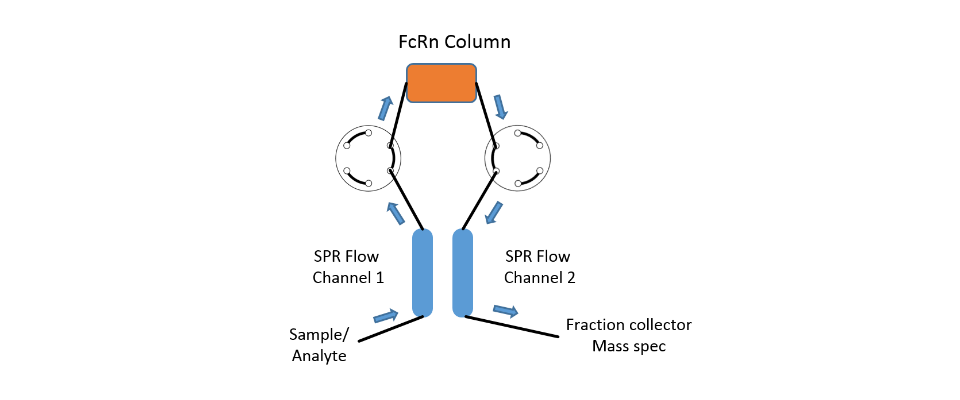
The SPR-PLUS system was equipped with the pneumatic pump, autosampler, the valve manifold incl. a µ-column and the fraction collector (Fig.1). Running buffer was PBS, pH 6.75 at a flow rate of 50 µL/min. The µ-column with a bed volume of 50 µL was filled with CM-Sephadex C-50, pre-equilibrated in running buffer overnight. The column was placed between the two flow channels of the 2SPR spectrometer (Fig. 2&3). The SPR sensor chip used was a CMD500L.

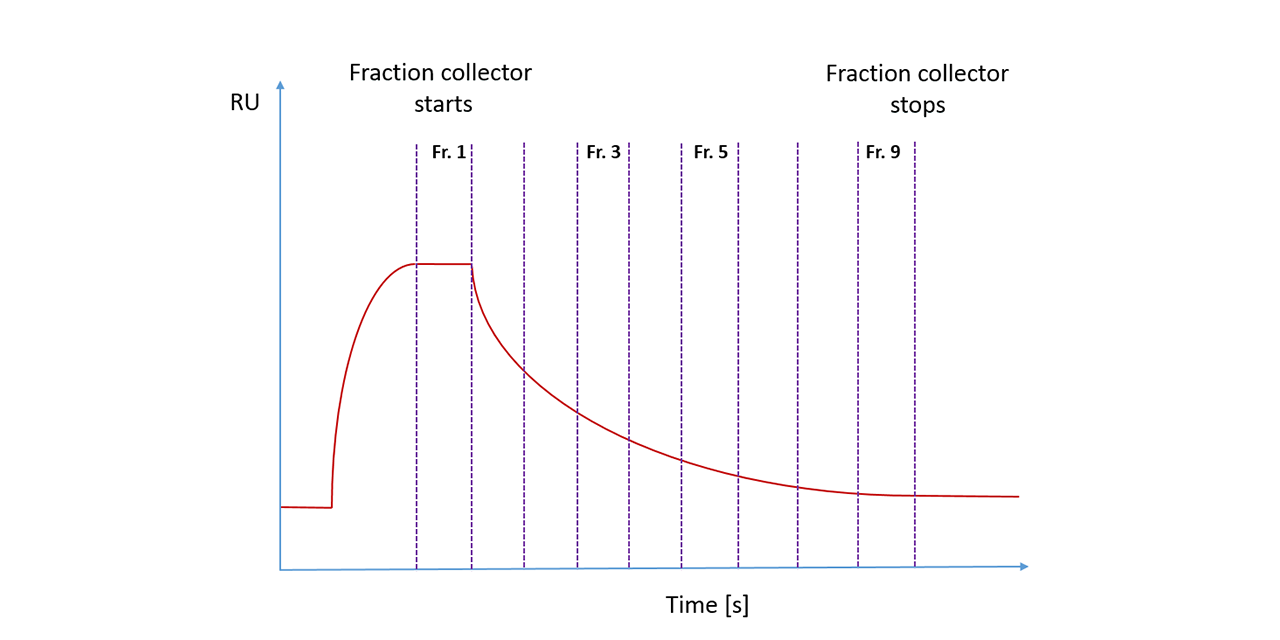
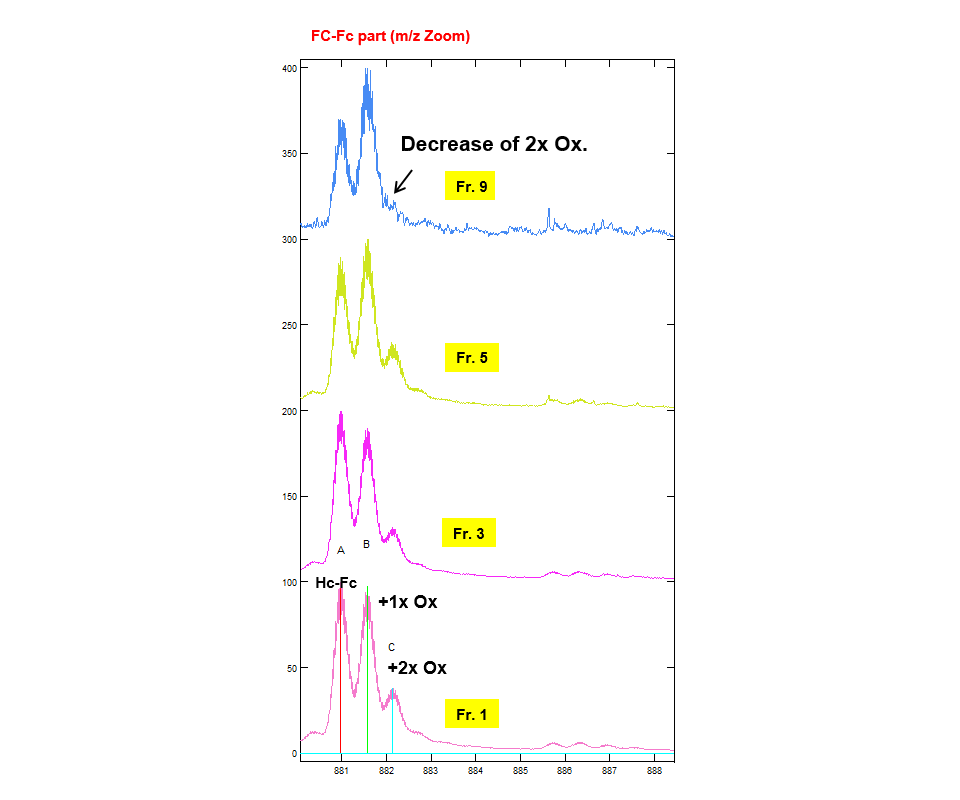
FcRn receptor was immobilized onto both flow cells and the µ-column by amine coupling with EDC/NHS. After immobilization of the receptor, unreacted NHS-esters were deactivated by 1M ethanolamine, pH 8.5. The antibody mixture (200 µg/mL) was injected at a flow rate of 100 µL/min for 3 min. Fraction collection started in the last minute of the association phase. Dissociation was carried out for 8 min (Fig. 4) with fraction collection. Each collected fraction was subsequently deglycosilated, IdeS digested, TCEP reduced, concentrated to a total volume of 60 µL and analyzed by UPLC-ESI-MS (Fig. 5).

Figure 2
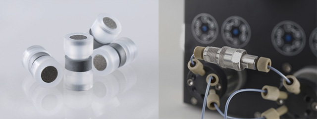
Figure 2: µ-column and µ-column holder mounted in SPR-PLUS valve manifold.
Figure 3
Figure 3: Schematic drawing of the fluidic pathway in the FcRn chromatographic application of the SPR-PLUS.

Results

SPR-PLUS analysis of the IgG-FcRn interaction of samples containing various antibody species differing in their affinity to the receptor has made it possible to resolve and quantify the individual molecular species in these samples. The new approach of combining SPR analysis with a miniaturized FcRn affinity chromatography is leading to lower sample consumption in the subsequent MS analysis step.

Note: SPR-PLUS supported FcRn chromatography only evaluates the structural and functional integrity of the antibody parts relevant for FcRn binding - it does not address the Fc receptors binding in general. Additional assays are required for a full evaluation of the functional integrity of antibody preparations. In fact, work is ongoing to establish a SPR-PLUS Fcγ receptor using affinity chromatography to further complement the functional analysis of antibodies.

Figure 4
Figure 4: Schematic sensorgram of antibody association and dissocation phase. Vertical lines indicating the collected fractions.
Figure 5
Figure 5: Mass spectra of the FC-Fc part from different fractions collected during the dissociation phase showing a decrease in the of the 2x oxidized variant of the antibody.

1. Schlothauer, T., Rueger, P., Stracke, J. O., Hertenberger, H., Fingas, F., Kling, L., ... & Koch, S. (2013, July). Analytical FcRn affinity chromatography for functional characterization of monoclonal antibodies. In MAbs (Vol. 5, No. 4, pp. 576-586). Taylor & Francis.

* The modules presented here have been developed as flexible additions to our range of SPR biosensors and serve merely as not binding application examples. Due to the variety of possible combinations and applications, we can not guarantee the suitability for a particular purpose, nor the freedom of third parties intellectual property. It is the responsibility of the end user to test the technical suitability of a setup and/or obtain eventually necessary licenses.

Application
option 02

SPR-PLUS as affinity chromatographic device: Investigation of pH-dependent dissociation of antibodies from their targets

Introduction

Chromatographers have been able to employ gradient separations for many years to obtain better separations and hence improved results. For instance, when running a chromatography experiment, one can employ a gradient to separate out impurites that might coelute when running with an isocratic buffer. The improved separation is typically based on changes in running buffer composition – eg. Increased percentage of organic solvent when using a reversed-phase column or increased polarity when using a normal phase column.

Now you can also use gradients to extend the range of information gained from each SPR injection. We offer a pulse free gradient pump (with degasser) that can be used to more efficiently and quickly determine analyte behavior. Users can vary such things as pH, salt content, detergent, and/or solvent content over the course of an analyte dissociation (or association or both).

SPR sample consumption is naturally lower than with chromtography, with typical flow rates being in the range of 25-100 μL/min.

Experimental

The SPR-PLUS system was equipped with the pneumatic gradient pump, autosampling unit and the 2SPR spectrometer. Running buffer was PBS, pH 5.5 at a flow rate of 50 µL/min. The SPR sensor chip used was a CMD500L.

FcRn receptor was immobilized onto flow cell one by amine coupling with EDC/NHS and the other flow was used as reference. After immobilization of the receptor, unreacted NHS-esters were deactivated by 1M ethanolamine, pH 8.5.

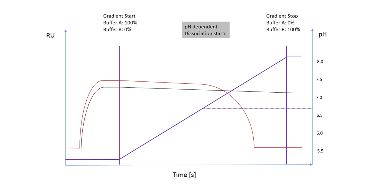
Both different engineered antibodies (5 µg/mL) were injected at a flow rate of 15 µL/min for 3 min. The gradient started after 2 min of dissociation going continuously from pH 5.5 to pH 8.0 in 20 min (Fig. 1).

Figure 1
Figure 1: Stacked SPR sensorgrams of association / dissociation behavior of two different engineered antibodies in a pH-gradient.

Summary

Addition of a gradient pump to an SPR setup provides added flexibility and utility. Use of a pulse-free pneumatic pump such as we employ here eliminates the effects of pump refills and provides accurate flow over a range of typical SPR flow rates. The ability to vary running buffer composition over the course of an injection can allow one to remove tight binders more gently than with traditional regeneration conditions and could allow users to selectively dissociate one binding component over another in a mixture where both bound to the same target. Since a fraction collector can easily be integrated into this setup, the effluent can then be taken to another detector (eg. a mass spectrometer) to identify what has been eluted.

Application
option 03

In depth characterization of binding parameters in stressed proteins by SPR and “gradient dissociation”

Figure 1
Figure 1: SPR-PLUS System comprising of a gradient pump, fraction collector and the universal and flexible valve manifold.

We show how the structural integrity of monoclonal antibodies of therapeutic interest (humanized , IgG1 class) can be analyzed with surface plasmon resonance (SPR) in depth. The interaction of structure-sensitive reagents with wild-type and several stressed variants of the IgG1s was studied by SPR using a gradient dissociation technique. Significant differences in binding patterns between wild-type and stressed variants of the IgG1s were revealed by the new SPR-PLUS concept which utilizes a gradient dissociation and which were not detectable by standard SPR alone.

This method is a valuable tool for monitoring structural integrity during upstream and downstream process development of therapeutic antibodies.

Materials and Methods

The Reichert SPR-PLUS system configuration is shown in Figure 1. The setup includes the standard components 2SPR spectrometer and autosampler along with a valve manifold and the unique SPR gradient pump.

For antibody characterization the sensor chip was immobilized with MabSelect protein at 20 µg/mL in 5 mM sodium formate buffer, pH 4.0. after activation with EDC/NHS chemistry (1% EDC in 100 mM NHS / 50 mM MES buffer NHS/MES buffer for 5 min). Subsequently residual active groups were quenched with 1 M ethanolamine, pH 8.5 for 4 min. As running buffer for the association phase and the initial dissociation phase PBS, pH 7.4 with 0.05% Tween P20 (PBST) was used. For running a gradient, a second buffer PBST at pH 2.0 was used. The gradient was performed from 0-100%. To ensure a full regeneration of both the MabSelect column and the sensor chip, 50 mM glycine HCl, pH 2.0 was injected for 4 min. The flow rate during the whole experiment was 20 µL/min. Wild-type antibody and force-stressed antibodies were injected at a concentration of 100 µg/mL with a contact time of 8 min. The gradient was started after 1100 sec after injection of each antibody sample and was carried out for 3600 sec.

Results

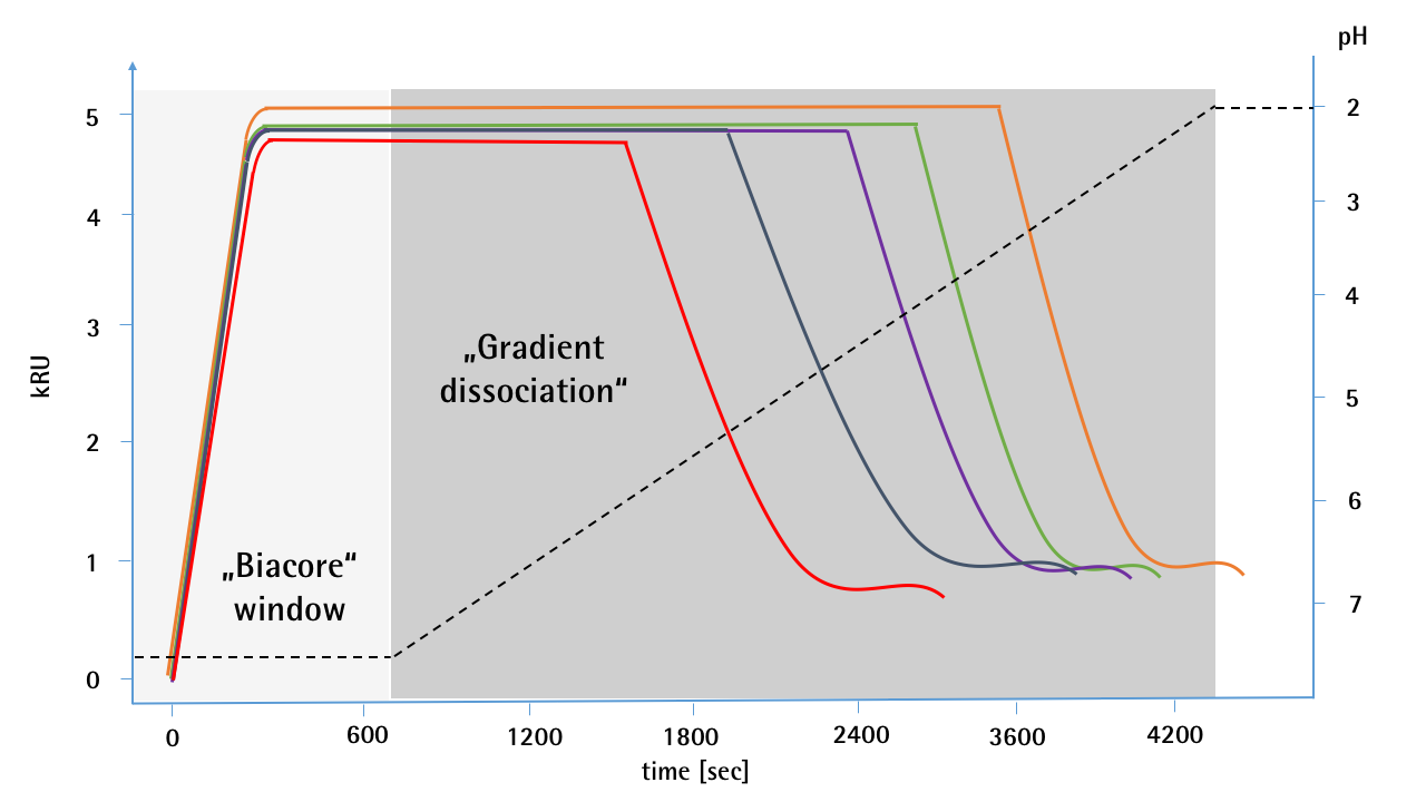
In the association phase and initial dissociation phase with typical durations for Biacore experiments only the wild type antibody showed a significantly different binding characteristic compared to the oxidized samples (1, 5, 10, 50 ppm H2O2) (Figure 2 “Biacore window”). It is also difficult to discriminate between the different states of oxidization. Only the 50 ppm stressed antibody seems to have a slight dissociation at pH 7.4.

It was much easier to distinguish between the individual samples when a pH gradient was carried out in the dissociation phase (Figure 2). Significant differences in the dissociation behavior lead to a clear distinction between the individual oxidation states of the antibodies.

Figure 2
Figure 2: Gradient dissociation sensorgrams of different forced oxidized monoclonal antibodies compared to unstressed wild-type. Subsequent to association and dissociation phases a gradient (PBST, pH 7.4 ➔ PBST, pH 2.0) was performed over 1h (red area) while in the normal association / dissociation only a difference between the unstressed and the stressed antibody was recognizable, the antibodies show marked changes during the gradient with increasing degree of oxidation in their binding behavior. Unstressed mAb = orange, 1 ppm H2O2 stressed mAb = green, 5 ppm H2O2 = green, 10 ppm H2O2 = violet, 20 ppm H2O2 = dark green, 50 ppm H2O2 = red.

Summary

Previous attempts to assay oxidative stress safely and with high resolution using SPR (Biacore) have often been unsuccessful. However, this limitation has been lifted by the innovative SPR-PLUS concept by XanTec / Reichert. Clearly and safely, even samples that had been exposed to comparatively low levels of oxidative stress could be distinguished from the non-stressed variant and samples with higher stress levels.

The new SPR-PLUS concept brings considerable advantages in terms of sample consumption, auto-automation and speed, especially for formulation development, in which the storage instability of biopharmaceutical products has to be tested.

Application
option 04

Miniaturized, referenced, SPR based self interaction chromatography (SPR-SIC)
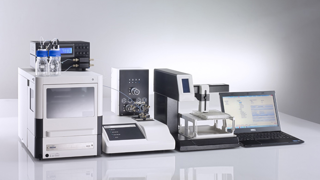
Figure 1

The use of proteins in industrial and pharmaceutical applications has become increasingly common. For example, a multitude of monoclonal antibodies (mAbs) are currently in biopharmaceutical drug development due to their high specificity and binding strengths. However, antibodies tend to aggregae and degrade at elevated concentration, and, as a consequence, lose their potency. In addition, protein drug formulations containing aggregates are known to cause an immunogenetic response and cannot be used. Preparation of very high concentrations of antibodies at a very low volume per dose is critical.

Therefore, predicting or quickly measuring aggregation/self-interaction behavior is preferred to have suitable formulations in the development of protein-based biopharmaceuticals [1].
Experimental methods for characterizing the influence of solution conditions (pH, ionic strength, etc.) would provide researchers with the opportunity to make predictions about how these variables will influence solubility, phase behavior and eventual crystallization. Protein-protein interaction phenomena occur throughout the full range of pharmaceutical protein-processing environments [2].

Protein-protein self-interactions under varying conditions are important, and as a screening tool to analyze these interactions self interaction chromatography (SIC) has been established for many years. But this method has the great disadvantage that very large amounts of protein are needed and the possibility for referencing is limited. With the fully automated surface plasmon resonance (SPR) based self interaction chromatography (SPR-SIC) these boundaries could be overcome.

SPR-SIC method
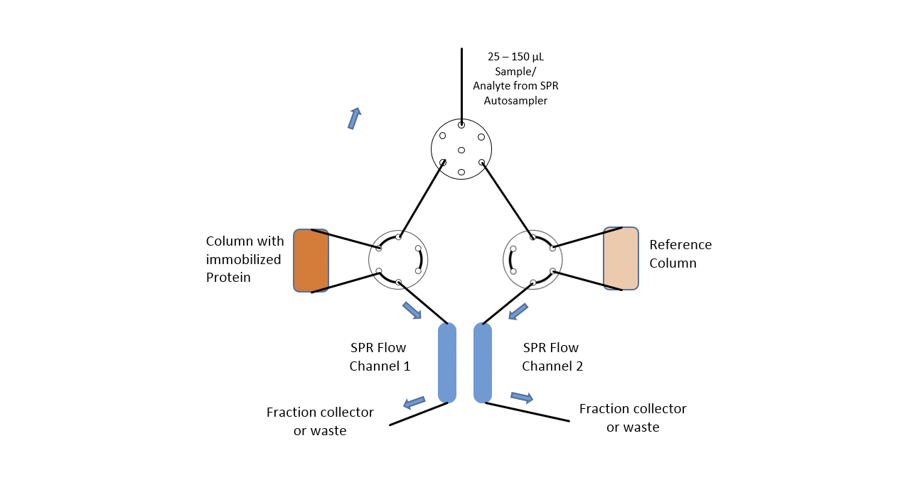
Figure 1

SPR, also known as the "Biacore" method, is widely associated with the determination of kinetics and active concentrations of proteins, peptides, and small drugs. Usually, these instruments are less flexible, closed black boxes that allow no other use or flexible design of experiments.
Not so, with the new and highly flexible SPR-PLUS concept, which was developed in collaboration with several leading pharmaceutical companies.

In this case, in addition to the high sensitivity of the SPR system, the extreme flexibility of the fluidics of the SPR-PLUS concept is exploited. Here, two μ-columns are integrated between the flow channels of the SPR system by means of an extended valve module, which serve as a reference and interaction column. Wherein the signal on the first SPR channel serves as a control for possible variations of the backpressure / packing density of the two columns and the second column downstream of the columns registers the retention time differences between the two columns.
The immobilization of the protein takes place automatically for the SPR flow channels through the autosampler. In the protein-loaded column, it is possible to choose between online immobilization and manual offline immobilization. If a calibration of the two columns is necessary this is done with injections of 1 M NaCl and 5% glycerol. The immobilization chemistry is dependent on the surface chemistries of both SPR sensor chip and used column material.

Materials and Methods

The system was equipped with two μ-column holders as shown in Figure 1. The μ-columns were filled with in each case 75 μL of CM-Sephadex 50 suspended in PBS. The column material for both columns was activated with 2% EDC in NHS / MES buffer (XanTec). For the protein column 100 μL of column material was washed several times with 5 mM sodium acetate pH 5.0 and then incubated with 200 μL of a 100 µg/mL mAb1 solution for 15 minutes. Subsequently, both column materials were quenched with 300 μL 1M ethanolamine, pH 8.5 to eliminate residual reactive groups.

Finally, the treated column materials were washed several times with PBS and then introduced into the columns. The antibody was immobilized online using a similar protocol on the sensor chip.
After incorporation of the column cartridges into the respective column holders, the entire SPR system was rinsed with 10 mM histidine as an air buffer. After equilibration of the columns and chip surface, different concentrations of the antibody (400, 200, 100, 50, 25, 12.5 μg / mL) were added for 4 min at 20 μL/min each over the protein and reference columns. Any non-specifically bound antibody was eluted by injections of 500 mM NaCl, 0.1 M Na borate, pH 7.8.

Results

The overlayed SPR sensorgrams (Fig. 2) show concentration dependent signals (peaks on the left side / refractive index jumps by concentration and non-specific binding) on the first SPR flow channel before the protein is in contact with the either the mAb-modified or reference column. The reproducibility of the injections for both flow paths (CH1-mAb-column-CH2 / CH1-reference-Column-CH2) indicates that both columns have the same backpressure. The peaks on the right side of the sensorgram (combination of refractive index by concentration and non-specific binding) show clearly different retention times between the reference and the mAb-modified column.
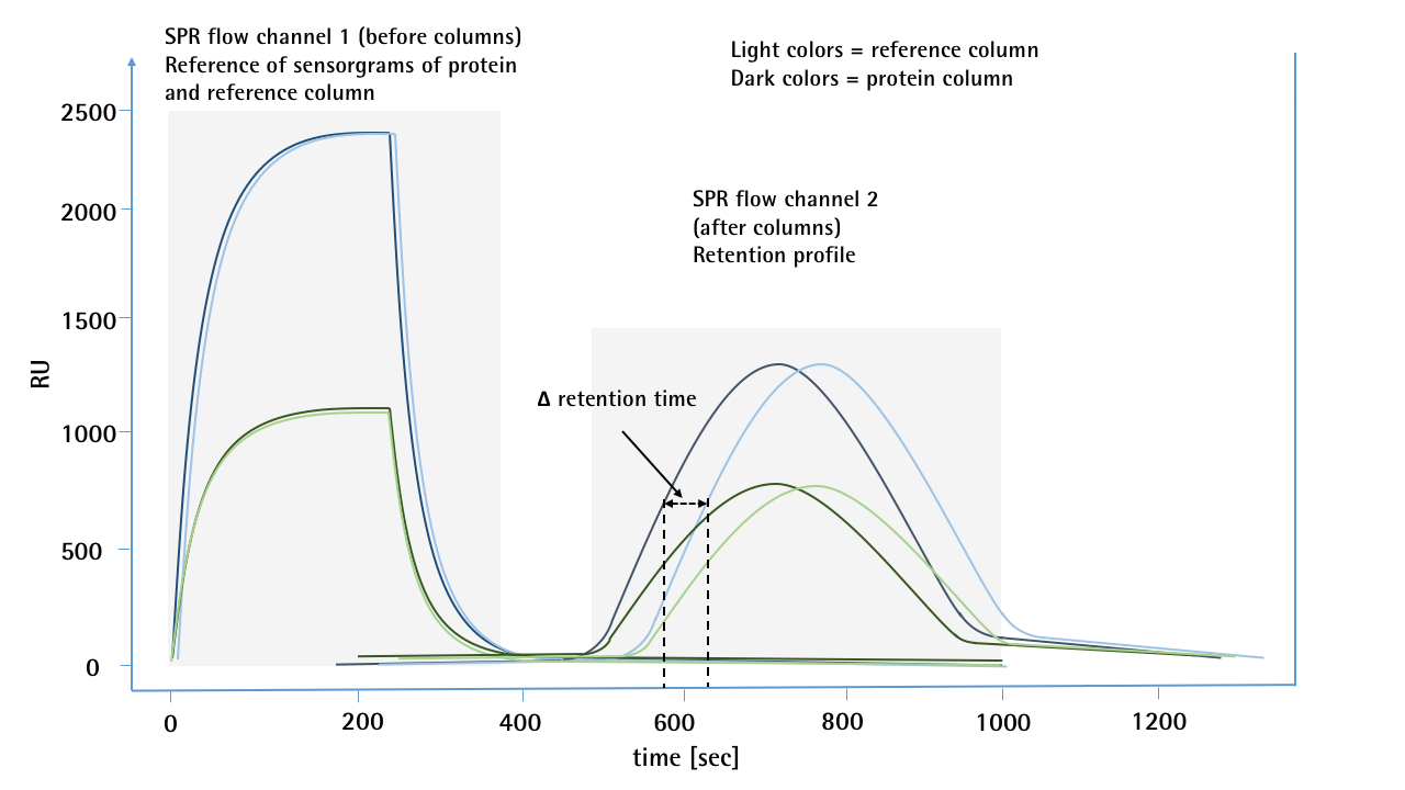
Figure 2
Figure 2: SPR sensorgrams of 2 different concentrations (10 & 50 µg/mL) of a mAb for self-interaction chromatography. For each concentration, a sample was first passed through the 1st SPR flow channel, then passed over a protein or reference column and then transferred to the 2nd flow channel. By means of salt injections, the different flow paths between the reference and the protein columns were previously calibrated and no deviation in the retention time was observed. The first flow channel of the SPR serves as a pre-column detector and shows no deviation for the two injections of one concentration, which would indicate any backpressure differences in the flow paths. The sensorgrams of the 2nd flow channel show a clear deviation in the retention time. In this case, the sample which was passed over the protein column was much more likely to reach the second flow channel than the sample which was passed over the unmodified reference column.

Summary and Conclusion

Understanding weak protein interactions is important for treating biological, crystallizing proteins, including for structure-based drug design, purifying protein mixtures, understanding protein diffusion in concentrated solutions, and stabilizing protein therapeutic formulations. However, these interactions are typically too weak to characterize in terms of quantities such as association constants that are suitable for strong protein interactions measured using methods such as surface plasmon resonance (SPR) [4]. Instead, of using SPR to characterize these weak protein interactions in the classic way, the SPR spectrometer was utilized as an ultra-sensitive detector for a miniaturized version of classical self-interaction chromatography (SIC) to characterize the interactions in terms of the osmotic second virial coefficient (B22). The new SPR concept significantly reduces the required amount of sample material and can therefore be located even in an earlier stage than in formulation development and also shows the application possibilities for other chromatographic questions.

  1. Deshpande, K. S., Ahamed, T., Ter Horst, J. H., Jansens, P. J., Van Der Wielen, L. A., & Ottens, M. (2009). The use of self‐interaction chromatography in stable formulation and crystallization of proteins. Biotechnology journal, 4(9), 1266-1277.
  2. Cleland, J. L., Powell, M. F., & Shire, S. J. (1993). The development of stable protein formulations: a close look at protein aggregation, deamidation, and oxidation. Critical reviews in therapeutic drug carrier systems, 10(4), 307-377.
  3. Ahamed, T., Ottens, M., van Dedem, G. W., & van der Wielen, L. A. (2005). Design of self-interaction chromatography as an analytical tool for predicting protein phase behavior. Journal of Chromatography A, 1089(1-2), 111-124.
  4. Tessier, P. M., Lenhoff, A. M., & Sandler, S. I. (2002). Rapid measurement of protein osmotic second virial coefficients by self-interaction chromatography. Biophysical journal, 82(3), 1620-1631.